the circulatory system.
heart dissection
Title Heart Dissection Prac Date Friday 25th October Partner Chelsea L
Aim To successfully dissect a heart in order to identify and understand the different parts and how they function to pump blood.
Materials A sheep's heart, a wooden board, newspaper, gloves, scissors, blade, forceps
External Examination
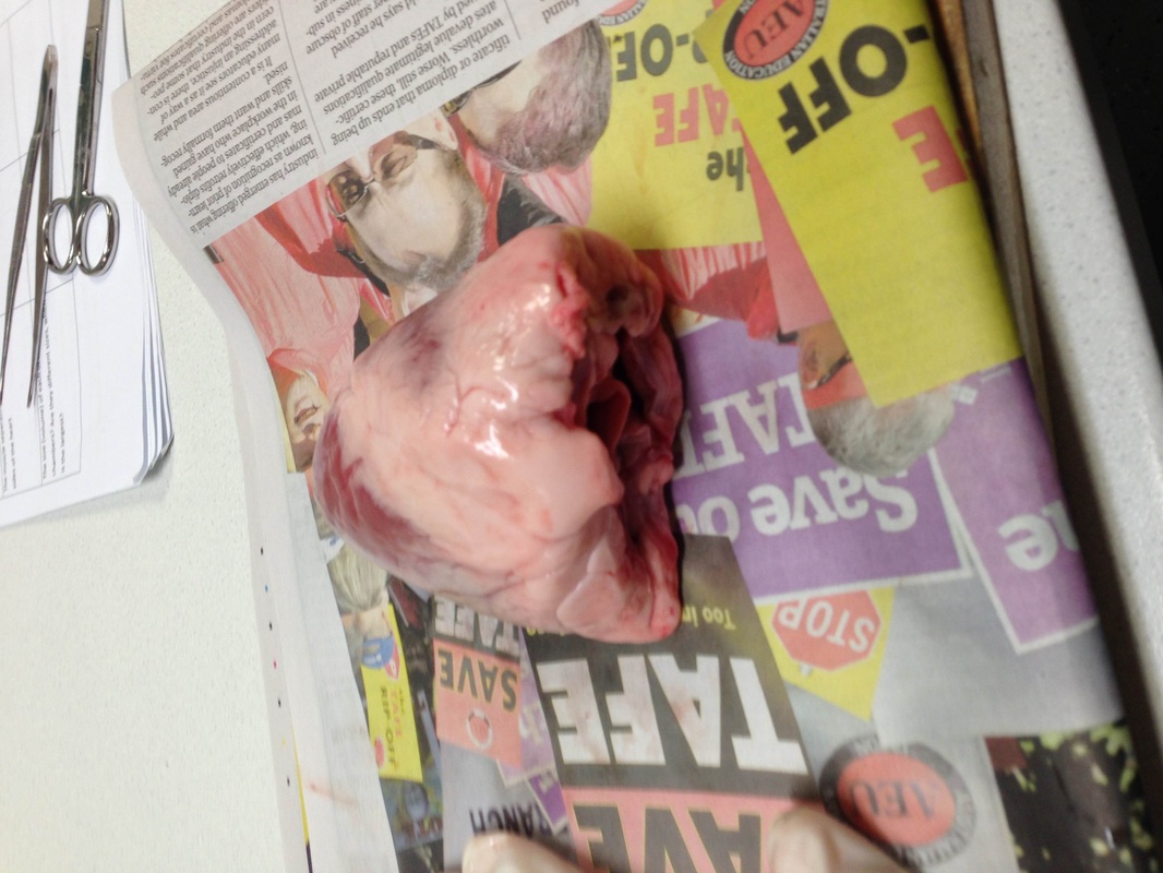
1. Describe the appearance of the heart. What did it look/feel like? What were some features you can describe?
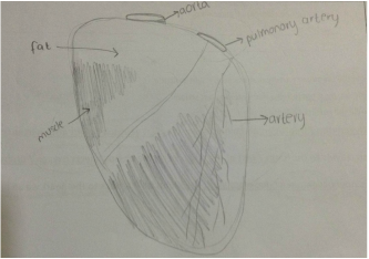
The heart was a reddish pink colour but there was a lot of white fat around it. The fat was very hard and crumbly. The heart felt muscular and thick. You could clearly see veins and arteries that look liked spidery thin lines along the surface. The aorta and the pulmonary artery were visible from the front exterior.
2. Provide a sketch of the front exterior of the heart and label all key parts visible.
See image to the left
3. Find and describe the blood vessels on the surface of the heart. (Coronary Arteries) And what would happen if they were blocked?
The coronary arteries were dark red/purple and the were very long and thin. They had lots of branches with smaller vessels attached. If they were blocked by a clot the heart would lack oxygen and and would have to work harder which may lead to a heart attack.
4. How do you know which is left and right on the heart?
The left ventricle which is on the left side has a thicker muscle as it has to pump blood upwards into the aorta. If you find this it's the left.
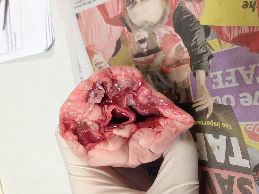
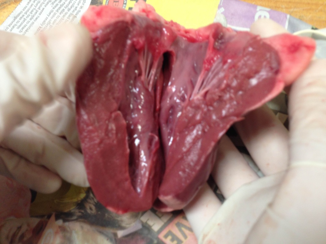
5a/b. Describe the difference between the thickness of the muscles at the top and bottom of the heart.
The muscles at the top of the heart feel thinner and a bit less dense. They are very elastic. The muscles at the bottom of the heart are harder and tougher. The feel dense and muscular.
5c. Describe the fat surrounding the heart.
There is a lot of fat surrounding the heart. It is very hard and an off white colour. The fat encloses the heart to protect it.
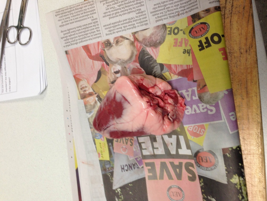
5d. Describe any major vessels entering and exiting the heart.
There were four that I could see- the vena cava, aorta, pulmonary artery and the pulmonary vein, The aorta and the pulmonary artery had tough elastic walls that were thicker than those of the vena cava and pulmonary vein.
6. Circle the correct answer.
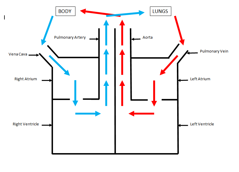
Deoxygenated blood leaves the right ventricle in an artery and travels to the lungs. Here the blood collects oxygen so it is now oxygenated. The blood travels back to the heart via a vein.
7a/b. Describe the thickness of the aorta. Why is it so thick? Where is it taking blood to?
The aorta carries blood from the heart to the brain and the rest of the body. It has a very thick elastic wall so it can pump the high pressure blood.
8a/b. Describe the thickness of the vena cava and compare it to the aorta. Which part of the heart does it go back into?
The vena cava has a thinner wall than the aorta because the pressure of the blood is not as high so it does not need to pump as hard. The vena cava re-enters the heart through the right atrium.
Internal Examination
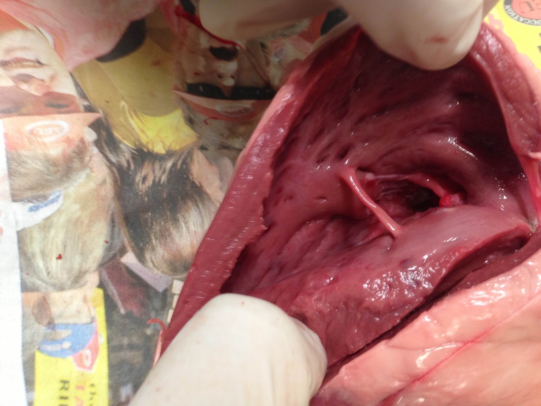
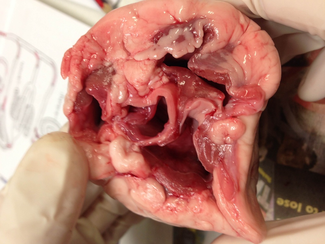
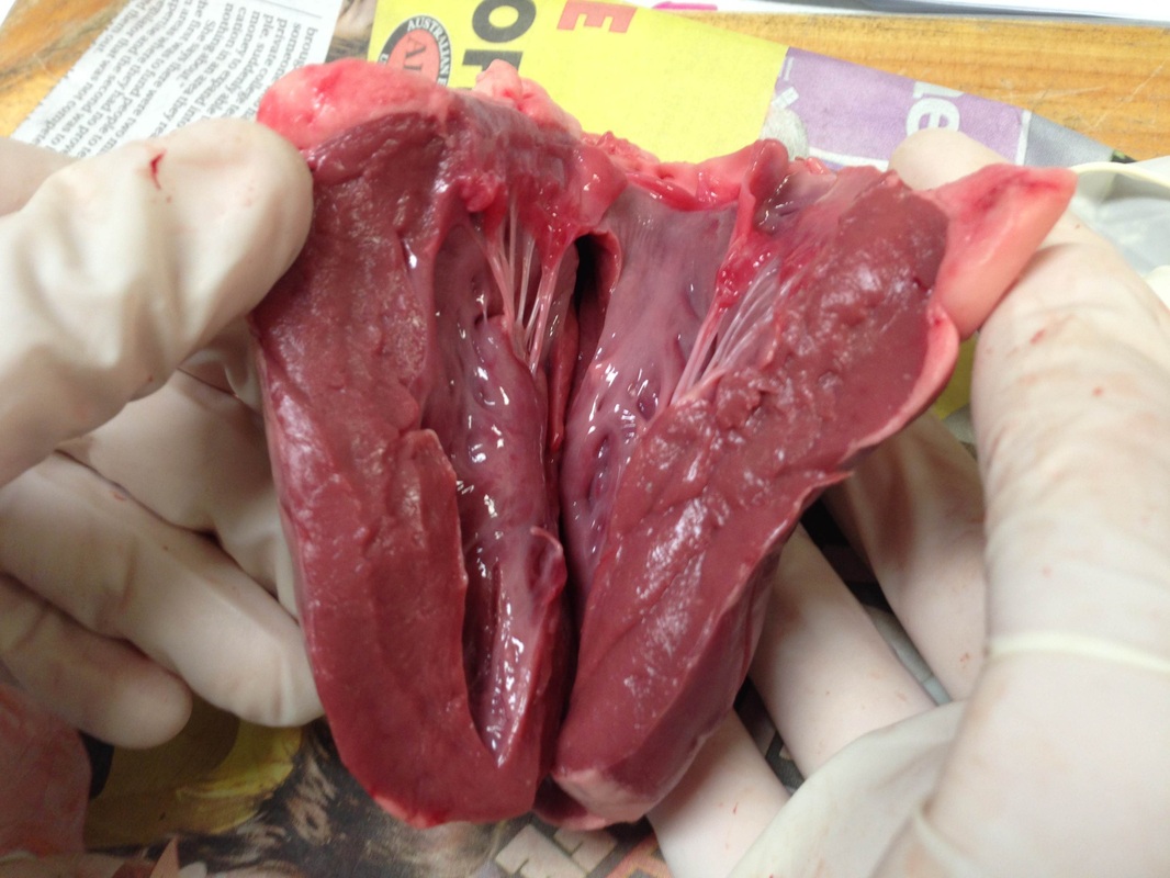
1. Describe what you see in the left side of the heart.
On the left side of the heart you can see the left ventricle and the left atrium. You can also see the top pulmonary vein and where it enters the left atrium.
2 Observe any valves you see. What might their job be?
There are valves separating the two chambers. They are to ensure blood flows in only one direction.
3. Cut the aorta. Describe how it feels and any other features.
Thee aorta has a thick wall and it was harder to cut than it looked. It was very tough and elastic and flexible. It was a lot wider than I thought!
Aim To successfully dissect a heart in order to identify and understand the different parts and how they function to pump blood.
Materials A sheep's heart, a wooden board, newspaper, gloves, scissors, blade, forceps
External Examination
1. Describe the appearance of the heart. What did it look/feel like? What were some features you can describe?
The heart was a reddish pink colour but there was a lot of white fat around it. The fat was very hard and crumbly. The heart felt muscular and thick. You could clearly see veins and arteries that look liked spidery thin lines along the surface. The aorta and the pulmonary artery were visible from the front exterior.
2. Provide a sketch of the front exterior of the heart and label all key parts visible.
See image to the left
3. Find and describe the blood vessels on the surface of the heart. (Coronary Arteries) And what would happen if they were blocked?
The coronary arteries were dark red/purple and the were very long and thin. They had lots of branches with smaller vessels attached. If they were blocked by a clot the heart would lack oxygen and and would have to work harder which may lead to a heart attack.
4. How do you know which is left and right on the heart?
The left ventricle which is on the left side has a thicker muscle as it has to pump blood upwards into the aorta. If you find this it's the left.
5a/b. Describe the difference between the thickness of the muscles at the top and bottom of the heart.
The muscles at the top of the heart feel thinner and a bit less dense. They are very elastic. The muscles at the bottom of the heart are harder and tougher. The feel dense and muscular.
5c. Describe the fat surrounding the heart.
There is a lot of fat surrounding the heart. It is very hard and an off white colour. The fat encloses the heart to protect it.
5d. Describe any major vessels entering and exiting the heart.
There were four that I could see- the vena cava, aorta, pulmonary artery and the pulmonary vein, The aorta and the pulmonary artery had tough elastic walls that were thicker than those of the vena cava and pulmonary vein.
6. Circle the correct answer.
Deoxygenated blood leaves the right ventricle in an artery and travels to the lungs. Here the blood collects oxygen so it is now oxygenated. The blood travels back to the heart via a vein.
7a/b. Describe the thickness of the aorta. Why is it so thick? Where is it taking blood to?
The aorta carries blood from the heart to the brain and the rest of the body. It has a very thick elastic wall so it can pump the high pressure blood.
8a/b. Describe the thickness of the vena cava and compare it to the aorta. Which part of the heart does it go back into?
The vena cava has a thinner wall than the aorta because the pressure of the blood is not as high so it does not need to pump as hard. The vena cava re-enters the heart through the right atrium.
Internal Examination
1. Describe what you see in the left side of the heart.
On the left side of the heart you can see the left ventricle and the left atrium. You can also see the top pulmonary vein and where it enters the left atrium.
2 Observe any valves you see. What might their job be?
There are valves separating the two chambers. They are to ensure blood flows in only one direction.
3. Cut the aorta. Describe how it feels and any other features.
Thee aorta has a thick wall and it was harder to cut than it looked. It was very tough and elastic and flexible. It was a lot wider than I thought!
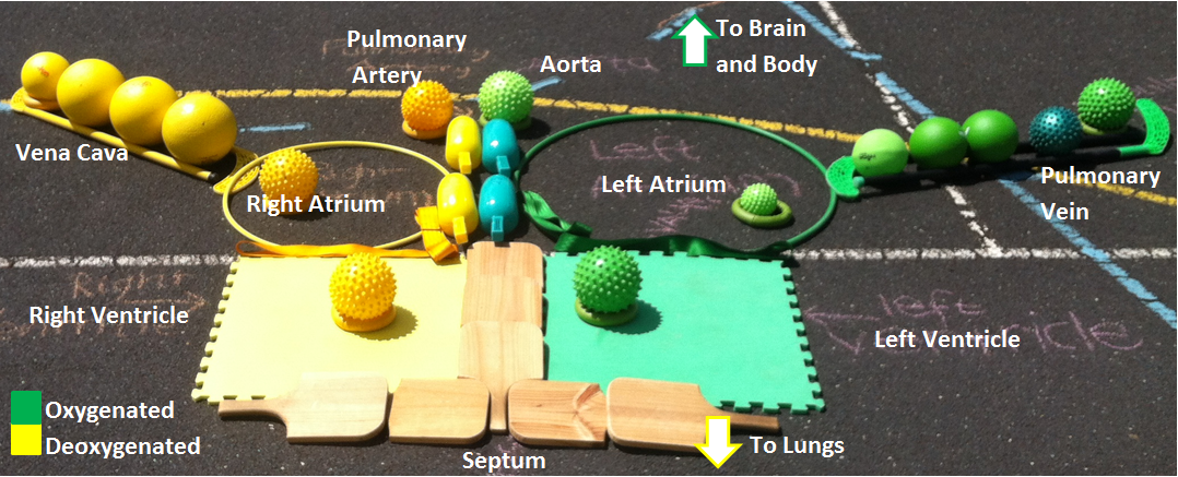
Using equipment from the sportshed we created our very own box heart diagram.
The oxygenated blood starts in the left ventricle (green mat) and is pumped out of the heart into the aorta (green shovels). The aorta takes the blood around to the brain and body where it is absorbed through capilliaries and becomes deoxygenated. This blood then returns to the heart via the vena cava (yellow balls) and into the right atrium (yellow hoop). The pulmonary artery (yellow shovels) takes the blood into the lungs where it is reoxygenated and returns through the pulmonary vein (green balls) back into the left atrium (green hoop).
The oxygenated blood starts in the left ventricle (green mat) and is pumped out of the heart into the aorta (green shovels). The aorta takes the blood around to the brain and body where it is absorbed through capilliaries and becomes deoxygenated. This blood then returns to the heart via the vena cava (yellow balls) and into the right atrium (yellow hoop). The pulmonary artery (yellow shovels) takes the blood into the lungs where it is reoxygenated and returns through the pulmonary vein (green balls) back into the left atrium (green hoop).