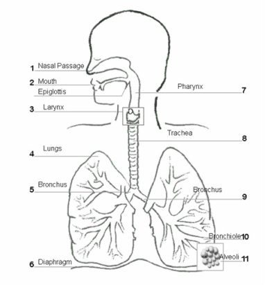
the respiratory system.
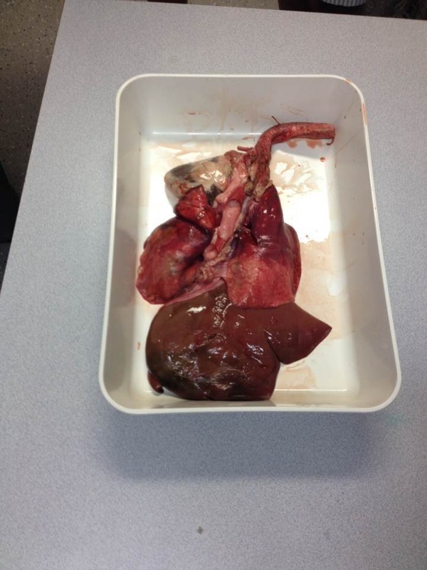
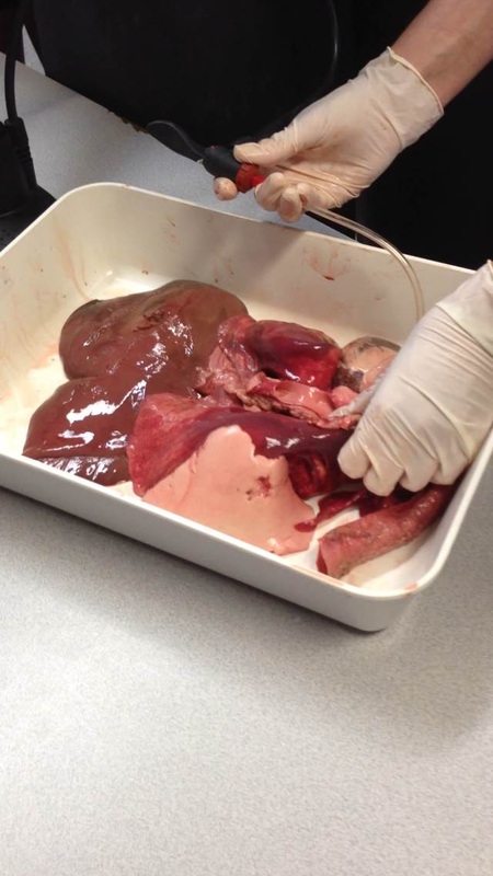
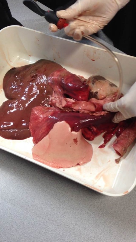
pluck demo
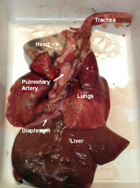
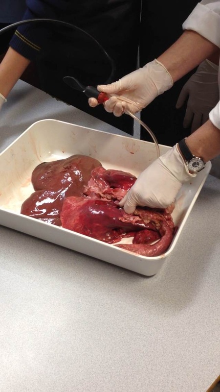
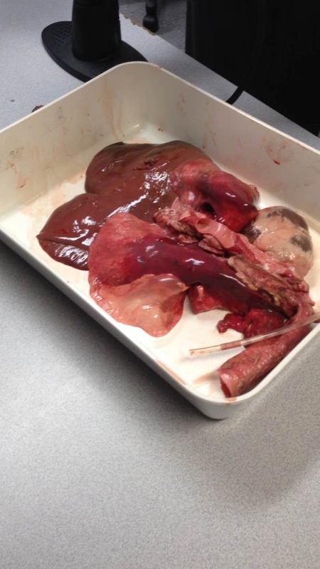
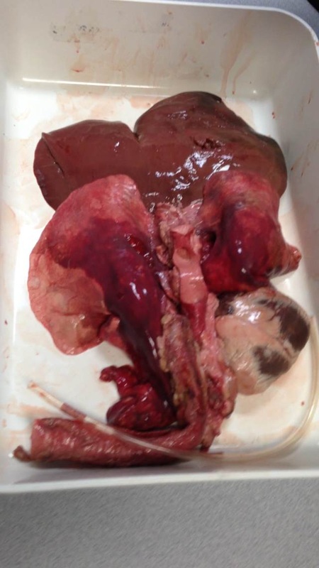
The pluck demo contained the heart, trachea, lungs and liver. Firstly we identified the trachea. We could feel the hard cartilage rings. Then we moved on to the heart. You could clearly see the aorta and vena cava, much clearer than when we did the heart dissection because it had not been cut the same way. The heart was connected by the pulmonary artery to the lungs. They felt very spongy and light. The pulmonary artery did not enter the lungs all at once but by many smaller entrances throughout both lungs. We attached the pump and blew air into the lungs. As they inflated they changed colour because they were becoming oxygenated! They did not inflate to their full size because they had a few small holes but they did grow in size a lot. There was a thin muscular layer at the bottom of the lungs which was part of the diaphragm. It was really tough and flexible. We had a quick look at the liver, it was hard and dense. It felt a bit like a tongue, and it was really big.